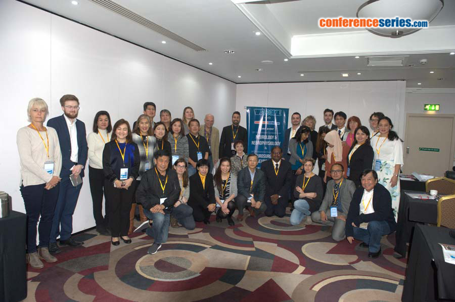
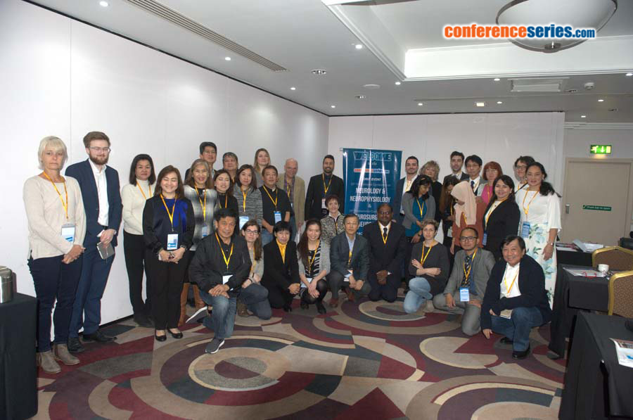
Day 2 :
Keynote Forum
Felix-Martin Werner
Medical doctor at the Euro Academy in Pößneck since 1999
Keynote: Agonists and antagonists of specific serotonergic receptors in the treatment of cognitive, depressive and psychotic symptoms in Alzheimer's disease
Time : 09:40-10:20
Biography:
Felix-Martin Werner studied Human Medicine at the University of Bonn. He has been working as a medical teacher in the formation of geriatric nurses, occupational therapists and assistants of the medical doctor at the Euro Academy in Pößneck since 1999. He has been doing scientific work at the Institute of Neurosciences of Castilla and León (INCYL) in Salamanca (Spain) since 2002. With Professor Rafael Coveñas, he assisted at over 30 national and 12 international congresses of neurology and published over 40 reviews and two books about neural networks in neurological and psychiatric diseases. Since 2014, he has belonged to the Editorial Board of the Journal of Cytology & Histology.
Abstract:
Alzheimer's disease is a neurodegenerative disease with cognitive, depressive and psychotic symptoms. We summarize the neurotransmitter alterations above all in the hippocampus and frontal/temporal cortices. In these brain areas, hypoactivity of acetylcholine and hyperactivity of noradrenaline at the beginning of the disease and hypoactivity of noradrenaline can be found. Glutamate exerts an excitotoxic effect and the presynaptic inhibitory neurotransmitter GABA shows hypoactivity. In depressive symptoms, a deficiency of monamines occurs in the brainstem and hippocampus. In psychotic symptoms, hyperactivity of dopamine and serotonin can be detected in the hippocampus and the ventral tegmental area. Neural networks in the corresponding brain areas are suggested. In Alzheimer's disease, 5-HT4 and 5-HT7 agonists and 5-HT3 and 5-HT6 antagonists have been suggested for the treatment of cognitive symptoms. In pioneer clinical studies, 5-HT4 agonists and 5-HT6 antagonists have shown a therapeutic effect, which was higher than placebo. In depressive symptoms, 5-HT reuptake inhibitors have a good therapeutic effect and can be used to treat aggressive behavior. In psychotic symptoms, 5-HT2A antagonists and second generation antipsychotic drugs can be administered, although the adverse effects should be considered. Because in Alzheimer's disease, a neurotransmitter imbalance between GABAA GABAergic neurons with hypoactivity and NMDA glutamatergic neurons with hyperactivity can be found, the first clinical studies with combined GABAA agonists and NMDA antagonists will be described.
Keynote Forum
Jinwei Zhang
University of Exeter, UK
Keynote: Activation of K+–Cl-–cotransporter KCC2 by inhibiting the WNK-SPAK kinase signalling as a novel therapeutic strategy for epilepsy
Time : 10:20:11:00

Biography:
Abstract:
The Cl--extruding transporter KCC2 (SLC12A5) critically modulates GABAA receptor signaling via its effect on neuronal Cl- homeostasis. Previous studies have shown that KCC2 was downregulated in both epileptic patients and various epileptic animal models. We discovered that the in vitro dual phosphorylation of Thr906 and Thr1007 in the intracellular carboxyl (C)-terminal domain of KCC2, mediated by the Cl--sensitive WNK-SPAK serine-threonine protein kinase complex, maintains the depolarizing action of GABA in immature neurons by antagonizing KCC2 Cl- extrusion capacity. GABAAR-mediated inhibition confines KCC2 to the plasma membrane, while antagonizing inhibition reduces KCC2 surface expression by increasing the lateral diffusion and endocytosis of the transporter. This mechanism utilizes Cl- as an intracellular secondary messenger and is dependent on phosphorylation of KCC2 at threonines 906 and 1007 by the Cl--sensing kinase WNK1. We propose this mechanism contributes to the homeostasis of synaptic inhibition by rapidly adjusting neuronal [Cl-]i to GABAAR activity. We further demonstrate here that this signaling pathway is rapidly and massively activated in an acute epilepsy model.This indicates that dephosphorylation of KCC2 at Thr906 and Thr1007 is a potent activator of KCC2 activity, and small molecular targets WNK-SAPK kinase signaling may be a novel therapeutic strategy for epilepsy.
- Neurology | Neurointensive Care Unit | Neurorehabilitation | Neurophysiology | Neurosurgery | Case Reports on Neurology & Neurosurgery | Skull base Neurosurgery
Location: Edinburgh, Scotland

Chair
Koji Abe
Okayama University, Japan

Co-Chair
Pedro Góes
Paulo Niemeyer State Brain Institute, Brazil
Session Introduction
David J Banayan
Rush University Medical Center, USA
Title: Advanced and emerging concepts in neuroprotection: Unlocking the secrets of nerve cell resilience and recovery

Biography:
David J Banayan MD, is an Assistant Professor in the Section of Psychiatry & Medicine at Rush University Medical Center. He is the Director of the Transplant Psychiatry Program, and Associate Director of Clinical Education for the Psychiatry Consultation Service. He is an board certified in General Psychiatry, Psychosomatic Medicine, and is a Fellow of the Royal College of Physicians of Canada. Following a Master’s degree in Clinical Epidemiology and Biostatistics at McMaster University, and Residency in Psychiatry at the University of Toronto, he completed a combined fellowship in Psychosomatic Medicine and Clinical Medical Ethics at the University of Chicago. His publications span the areas of intensive care medicine, deliruim, adolescent suicide, first episode psychosis, and research ethics. His areas of current academic interest include quality improvement in transplant psychiatry, neuroprotection, and the complex interface between general medicine, psychiatry, ethics, and the law.
Abstract:
Neuroprotection is a burgeoning area of scientific research. Certain pharmaceuticals and nutraceuticals have the potential to modify and enhance nerve cell response to toxic stimuli. This discovery has spawned intense interest in unlocking the cellular mechanisms that confer such resilience and recovery. Numerous biochemical pathways play a role in neuroprotection, such as: enhanced neutralization of molecular radicals; mitochondrial membrane integrity support; arresting generation of pro-inflammatory cell membrane metabolism products; activation of neurotrophic factors; modification of intracellular calcium homeostasis; inducing shifts in the resting endogenous balance of pro-apoptotic and anti-apoptotic factors within the cell; and others. This session introduces participants to fundamental and advanced concepts in neuroprotection through an examination of the downstream mechanisms of psychotropic agents and nutraceuticals. Recent advances in neuroprotection are also reviewed. The session will prepare clinicians to engage the literature on neuroprotection with an informed, critical eye. Proprietary animations developed by the author, bring to life difficult-to-understand abstract concepts, and provide a unique learning experience for participants. Comprehensive critical review of the biochemical sciences and biomedical literature through PubMed and EMBASE. While bench research and animal studies currently dominate the neuroprotection literature, as this nascent area of science evolves, it is hoped that it will culminate in the development of specific sub-cellular targets in humans. Human studies are costly, complicated, and require a large number of participants to show an effect, posing a potential barrier for real-world progress in neuroprotection.
Jacintha Vikeneswary Francis
Hospital Sultanah Nur Zahirah, Malaysia
Title: A challenge in managing posterior fossa cyst with hydrocephalus: A case report
Biography:
Jacintha Vikeneswary Francis is presently the Neurosurgeon, Ministry Of Health Malaysia, Hospital Sultanah Aminah Johor bahru, Malaysia since June 2016. She served as Neurosurgical Medical Officer in Hospital Kuala Lumpur and Hospital Queen Elizabeth, Kota Kinabalu Sabah from November 2006 till 2012. She attended several Conferences and was Chairman for few.
Abstract:
Background: Posterior fossa cysts are benign but usually developmental lesions. There are patient usually asymptomatic. Some cysts might communicate with these subarachnoid spaces however non-communicating cyst could develop from communicating ones giving rise to complexity of diagnosis and further management.
Case Description: One year old baby girl presented with developmental delay which is gross motor and speech with increase ICP symptoms. CT brain and MRI revealed large posterior fossa cyst extending supratentorially causing compression to the third ventricle and showing gross hydrocephalus. We proceed with endoscopic fenestration of posterior fossa cyst and ommaya insertion and subsequently proceeded with ventricular peritoneal shunt. Due to previous history of necrotizing enterocolitis (NEC), patient developed malfunction of the shunt due to poor absorption of the shunt, eventually patient required ventriculo-cysto-atrial shunt. 6 months post-procedure, patient currently does not show any evidence of recurrent cyst and developmentally showing improvement.
Conclusion: Posterior fossa cyst is rare and contributes to challenges radiologically and surgically. Current surgical management mainly depends more on clinical features giving little insights on the exact pathology of posterior fossa cyst.
Tiziana Bonifacino
Tiziana Bonifacino PhD, is an Assistant Professor at the Department of Pharmacy, University of Genoa, Italy.
Title: Excessive glutamate release and undelying synaptic mechanisms in a mouse model of amyotrophic lateral sclerosis.

Biography:
She got the degree in Chemistry and Pharmaceutical Technology (honors) in 2007 and the PhD in Neurochemistry and Neurobiology in 2011. The present scientific interests are related to neuronal transmission in the CNS and are focused on the cellular and molecular mechanisms of neurotransmitter release and receptor activity in physiological and pathological conditions, such as amyotrophic lateral sclerosis. She has established several scientific collaborations with national and international institutions. She has published 32 publications (25 papers, 6 abstract on journals and 1 graphical abstract) in peer-reviewed journals
Abstract:
Amyotrophic lateral sclerosis (ALS) is a fatal neurodegenerative disease, characterized by upper and lower motor neuron degeneration. Glutamate(Glu)-mediated excitotoxicity plays a major role in cell death and Glu has been found elevated in serum of patients and animal models of ALS. The aim of this research was to investigate whether the augmentend extracellular Glu can be sustainend by alteration of its release. Glu release was studied from purified spinal cord synaptosomes of SOD1G93A mice at the early and the late phases of the disease (4 and 17 weeks of life, respectively). Both the spontaneous and the stimulus-evoked exocytotic Glu release were increased at the two stages studied. We also measured the expression/activation state of a number of pre-synaptic proteins involved in neurotransmitter release: few of them were found modified and synaptotagmin and actin resulted over-expressed in both 4 and 17 week-old mice. Increased pre-synaptic Ca2+ levels, over-activation of calcium/calmodulin-dependent kinase-II and ERK/MAP kinases correlate with hyper-phosphorylation of synapsin-I at both stages. In line with these findings, Glu exocytosis was paralleled by the increase of the readily releasable pool of vesicles and prevented by blocking synapsin-I phosphorylation, using specific antibodies. Our results highlight that abnormal glutamate release is present in the spinal cord of SOD1G93A mice at the pre-symptomatic and late stage of the disease, an event accompanied by marked plastic changes of specific pre-synaptic mechanisms supporting exocytosis, that in turn may represent targets to diminish exitotoxicity in ALS. The precociousness of this phenomenon may imply that it represents a cause rather than a consequence of the neuronal damage during disease progression.
Itaru Yazawa
Lecturer at Hoshi University School of Pharmacy and Pharmaceutical Sciences since 2015
Title: A study on functional interactions between the central nervous system using a decerebrated and arterially perfused in situ preparation

Biography:
Itaru Yazawa has been a Visiting Lecturer at Hoshi University School of Pharmacy and Pharmaceutical Sciences since 2015. He received his PhD in Physiology from Tokyo Medical and Dental University in 2002. With using the decebrated and arterially perfused preparation he developed, he found that there are autonomous reciprocal functional interactions between the brainstem and the spinal cord. The preparation is recognized as a vital tool for investigating functional interactions between the CNS in several research fields. Currently, he is focusing on constructing a new system where the preparation can be maintained at the same body temperature as living animals.
Abstract:
Most recent studies of motor behavior use in vitro preparations of the neonatal rodent brainstem-spinal cord and spinal cord. However, the relevance of these studies to the neural mechanisms of adult respiration/circulation and locomotion is unclear, because the neonatal brainstem and spinal cord are immature. Moreover, it has been reported that the oxygen tension in deep tissues in these preparations was extremely low compared to that in the in vivo preparations. Thus, the results obtained from these in vitro preparations might indicate the physiological phenomena produced in a hypoxic state. To overcome these difficulties in research fields using rodents, we have adapted a decerebrated and arterially perfused in situ preparation originally developed by Pickering et al., to the preparation that can investigate functional interactions between the central nervous system. For this purpose, the rodent (weighing about 5 g or more) was decerebrated and survived by a type of total artificial cardiopulmonary bypass as a means of extracorporeal circulation to deliver oxygen to the tissues of the entire body. The oxygen and ion components of body fluid required for the survival of the preparation were supplied via the blood vessels. The physiological state of this preparation resembles to that of unanesthetized rodents under hypothermia. Here we would like to introduce a decerebrated and arterially perfused in situ preparation, and talk about reciprocal functional interactions between the brainstem and the spinal cord found by using this preparation.
Koji Abe
Professor and Chairman of Neurology at Okayama University Medical School in Japan
Title: Neuroprotective therapy with antioxidative drugs and supplements

Biography:
Koji Abe is currently a Professor and Chairman of Neurology at Okayama University Medical School in Japan. He graduated MD from Tohoku University School of Medicine, and then got PhD title from Tohoku University under direction of Professor Kyuya Kogure. He has been publishing more than 350 papers on cerebral blood flow and metabolism and neurodegenerative diseases. His research interests cover many important fields of neurology especially in the mechanism of ischemic brain damage, gene and stem cell therapy, neuroprotection, and neuroimaging. He is the Past President of the International Society of Cerebral Blood Flow and Metabolism (CBFM), and organized World CBFM meeting in Osaka in 2007 and Japan-Asia CBFM meeting Okayama city in 2014. He is currently serving Presidents of both Vas-Cog Japan and Vas-Cog Asia societies.
Abstract:
Neuroprotection is essential for potential therapy not only in acute stage of stroke but also in chronic progressive neurodegenerative diseases such as ALS, Parkinson’s disease (PD), and Alzheimer’s disease (AD). Free radical scavenger can be such a neuroprotective candidate with inhibiting death signals and potentiating survival signals under cerebral ischemia and even neurodegenerative cellular processes. Edaravone is one such free radical scavenger, which is the first clinical drug for neuroprotection in the world and has been used from 2001 in most ischemic stroke patients in Japan. A recent multicenter prospective double-blind placebo-control clinical trial with edaravone for ALS patients conducted in Japan showed a positive effect for delaying the clinical score (ALSFRS-R) during the course of examination (24 weeks). Serious or critical adverse effect was not noted in this clinical trial. Of particular was that this clinical benefit of edaravone was shown as an add-on therapy after anti-glutamatergic riluzole. These data strongly suggest a potential underlying mechanism of oxidative stress in ALS and a clinical delay by a free radical scavenger. Antioxidative supplements are also important choices to prevent or even treat neurological disorders such as ischemic stroke and dementia. Many basic and clinical studies suggest that antioxidative supplements such as Tocovid® (mainly tocotrienol) and TwendeeX® (coenzyme Q+multivitamin) may be effective to ameliorate acute ischemic stroke, chronic cerebral white matter damage, and Alzheimer’s dementia.
Ekaterina Drozhevskaia
Clinical Neurophysiologist in Turner Scientific Research Institute for Children's Orthopedics
Title: Complex clinical and functional study of children with idiopathic tip-toe walking

Biography:
Ekaterina Drozhevskaia graduated from St. Petersburg State Pediatric Medical Academy in 2008, finished Residency in Clinical Neurophysiology at North-Western State Medical University in 2014. She was Head of Diagnostic Department in Institute of Human Brain, St. Petersburg, Russia and now working as Clinical Neurophysiologist in Turner Scientific Research Institute for Children's Orthopedics and is Chairman of Neuromuscular Disorders Society in St. Petersburg from 2015.
Abstract:
Tip-toe walking is a well-known problem in children which leads their parents to consult a specialist. This condition usually is difficult to differentiate between neurological and orthopedic disorder that’s why it needs a multidisciplinary approach and cooperation of different specialists to find a diagnosis and determine the correct treatment. 25 children with tip-toe walking (aged 1-15 years) were examined by neurologist and orthopedist. To find out the main reason of walking disorder a number of neurophysiological and radiodiagnostic methods were used. The neurophysiological investigations included EMG of lower limb muscles, NCS, TMS. Imaging included US and MRI of lower limbs muscles. Radiological method was chosen according to the age of a child. Based on the results of all examinations and investigations children were put to a different group: CNS disorder, neuromuscular disorder, orthopedic disorder - and they got treatment accordingly. These results show the importance of considering not only clinical examination by specialists, but also different types of investigations (neurophysiological and imaging) for optimizing the approach to full diagnostic and proper treatment and rehabilitation approach to children with idiopathic tip-toe walking.
- Clinical Neurophysiology | Neuronal functions and disorders | Neurorehabilitation| Neuroimaging | Spinal Surgery | Neurosurgery | Brain Tumour
Chair
Felix-Martin Werner
Euro Akademie Pößneck, Germany
Co-Chair
Tamajit Chakraborty
Sir Ganga Ram Hospital, India
Session Introduction
Ludmila Zylinska
Medical University of Lodz, Poland
Title: Neuronal calcium dyshomeostasis as a crucial process in neuropathologies
Biography:
Ludmila Zylinska PhD, DSc, is a Full Professor and Head of the Department of Molecular Neurochemistry, Medical University of Lodz, Poland. Her scientific interests are related to regulation of neuronal calcium homeostasis with particular attention brought on plasma membrane calcium pumps in the CNS, as well as on molecular mechanisms of calcium-dependent processes that regulate neuronal transmission in physiological and pathological conditions. She has published more than 60 papers in the areas of Biochemistry, Neuroscience and Cell Biology.
Abstract:
Oscillations of cytosolic Ca2+ are necessary for cellular signaling and propagation of Ca2+ signal is an absolute requirement for the functioning of neuronal cells. Ca2+ appears to be a universal and ubiquitous signaling molecule, thereby in resting neuronal cells its concentration is kept at ~100 nM against 1-2 mM outside the cell. Inability to maintain calcium homeostasis in neurons underlies many neuropathologies. Ca2+ may elicit a variety of different responses based on the type of targeted neurotransmission pathways, and it regulates synaptic plasticity, controls neuronal growth and neuronal survival. The plasma membrane contains a high affinity Ca2+-ATPase (PMCA) that translocates Ca2+ from the cytosol to the extracellular environment. The enzyme is coded by four separate genes (PMCA1-4), among which PMCA2 and PMCA3 are considered as neuron-specific forms. In the brain, PMCA function declines progressively during aging, thus impaired calcium homeostasis may contribute to neurodegeneration. We have developed the stable transfected differentiated PC12 cells with reduced level of PMCA2 or PMCA3, and the most critical finding was permanently increased resting Ca2+ concentration. Altered PMCA composition affected the expression level of several Ca2+-associated proteins (SERCA, calmodulin, calcineurin, neuromodulin) and certain types of voltage-gated calcium channels. We have also evidenced a novel PMCA role in regulation of bioenergetic pathways and mitochondrial activity. Interestingly, some changes could occur as adaptive processes protecting cells against calcium overload. Since age-related PMCA decrease has been documented, our modified PC12 cells may be a useful model to clarify the biological changes in neurons as well as to study the vulnerability of cells to neurodegenerative insults.
Jaideep J Rayapudi
Pondicherry Institute of Medical Sciences, India
Title: Challenges to human intelligence in a future with artificial intelligence

Biography:
Dr. Jaideep J Rayapudi, completed MD Physiology in 2003 and has been involved in training & development of Medical Students with a keen interest in the process of learning & memory. He has been at the cutting-edge of developing & implementing technology-based education systems and advocates greater use of artificial intelligence to improve healthcare and healthcare education. He believes that the only way to know the future is to build it and that begins by challenging the status quo.
Abstract:
A lot of discussion abounds today about the future of Humanity, society and community with respect to the advancing prospects of all-pervasive artificial intelligence in the coming days; be it the voice activated devices, self-driven cars, financial trading or even robot-driven healthcare. In the background of all these massive and quick changes how does the human brain respond & react? The industrial revolution took away jobs which had traditionally required human muscle power and now in the information age we are poised to lose out jobs to machines where Human Intellectual work is required. Outside of the job space too, much of decision-making and thought has been aided heavily by data driven machine learning algorithms.
Neuroplasticity can enable the brain to get better and learn more; can the opposite be true with disuse of critical components of our cognitive behaviour? The future challenge for cognitive neuroscientists would be to guard & guide brains with suitable strategies to minimise the damage due to the onslaught of machine intelligence. Steps must be taken to recognise relevant issues and diagnose & intervene wisely globally. While apocalyptic scenarios like in the Terminator / Matrix may be prevented by sound policy; neural responses and its subsequent effects in areas of Psychology, society, polity, culture, art, design, relationships, religion etc must be prepared for and countered / augmented if needed. This proposed talk seeks to highlight some of these concerns and generate dialogue, response and action in this matter.
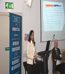
Dharitri Parmar
Government Medical College, Surat, India
Title: Review on omega fatty acids and neurophysiology
Biography:
Dharitri Parmar is working as Professor in Physiology since 2007 and as Teacher in medical college since 1992. She has contributed almost 22 years as teacher is in Government Medical College, Surat, India which is affiliated to Veer Narmad South Gujarat University. She is multifaceted personality who is interested in administration and academic activities. Currently holding the post of Addittional Dean of the Government Medical College. She has worked on few reserch topics and floated presentation online. She has visited nearly 50 medical schools in India and overseas as assessor (deputed by Medical council of India) : as an examiner or as an interviewer. She has also reviewd number of publications for journals.
Abstract:
Omega 3 and 6 are different types of PUFA having distinct effects on human systems. EFA is most debatable and important nowadays while studying nutritional role of lipids and its effects in physiology. This oral presentation is to focus on recent advances in importance of ratio (omega 6: omega 3) for clinical purposes physiologically. Omega 3 and Omega 6 compete for enzymes to produce signaling molecules, to modulate gene expression and to consolidate cell-membrane. The ratio of omega 6 to omega 3 is of importance according to various studies and its effects on nervous system physiologically, which may differ for different tissues or functions. Many hypothesis and various mechanisms are discussed herewith in context to BBB, membrane fluidity, myelin sheath, neurotransmitters etc. As a researcher it is studied for physiology of Learning, memory, sleep, early development, aging, and stress with EFA. Results conclude that 4:1 ratio has protective and stabilizing effects on neuron and its functions.The ratio may be a key factor in modulating behavioral, developmental, pharmacological effects of lipids.

Biography:
Rebecca B Baker holds two Bachelors degrees: one in Nursing from the University of Tennessee, the Health Science Center in Memphis and one in Education from the University of Memphis. Currently she is completing pre-requisites to purue a Doctor of Nursing Practice. She serves as the Chief Clinical Officer at Utilize Health, a population health compamy that specializes in care management services for patients with severe neurological conditions. She is a veteran nurse with over twenty-six years of clinical experience working with patients and care teams in a variety of settings.
Abstract:
Why aren’t long term post acute outcomes and recovery documented in patients with neurological conditions such as severe spinal cord injury (SCI)? As a healthcare system, how far should we reach in documenting outcomes and recovery and for what length of time so that current knowledge aligns with rehabilitation approaches for mobility recovery? Historically, research for the severe SCI patient population has looked at smaller sample sizes and the patients are less than 1-2 years post injury or diagnosis. In this presentation, we explore what patient reported outcomes can do to shift the conversation around what mobility recovery and tarteged rehabilitation therapies in SCI looks like. A key factor in moving the conversation regarding expected outcomes requires a fundamental hypothesis that recovery from severe spinal cord injury is a life-long process. Therefore, with research and data collection at scale (tens of thousand of patients), we can start to tie causation to pieces of recovery and even rehabilitation timelines and therapies that may not have been considered in the past. Case studies will be presented and explores a sample case: a 17 year old sustained an Asia A complete SCI at C6-T5. After nearly 6 years, she learned to walk unasissted. Even after 14 years (she is 31 today), she continues to recover. Collecting and analyzing patient reported outcomes at scale can shed light on long term recovery and what is possible in a poulation that has historcially been given little to no hope in mobility recovery.
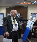
Felix-Martin Werner
Euro Akademie Pößneck, Germany
Title: Update of the neural networks involved in generalized epilepsy
Biography:
Felix-Martin Werner studied Human Medicine at the University of Bonn. He has been working as a medical teacher in the formation of geriatric nurses, occupational therapists and assistants of the Medical Doctor at the Euro Academy in Pößneck since 1999. He has been doing scientific work at the Institute of Neurosciences of Castilla and León (INCYL) in Salamanca, Spain since 2002. With Professor Rafael Coveñas, he assisted at over 30 national and 12 international Congresses of Neurology and published over 40 reviews and two books about neural networks in neurological and psychiatric diseases. Since 2014, he has belonged to the Editorial Board of the Journal of Cytology & Histology.
Abstract:
We reviewed the alterations of neurotransmitters and neuropeptides in the following brain areas involved in generalized epilepsy: hippocampus, hypothalamus, thalamus and cerebral cortex. In these brain areas, neural networks are also actualized. The mechanisms of action of newer antiepileptic drugs, for example a GABAB agonist, an AMPA receptor antagonist and brivaracetam, used in the treatment of generalized epilepsy are also discussed. Updating the neural networks, we suggest that in the hippocampus GABAergic neurons presynaptically inhibit, via GABAB receptors, epileptogenic neurons. GABAergic, glutamatergic, serotonergic and dopaminergic neurons form the principal neural network, while GABA and serotonin deficiency and dopamine and glutamate hyperactivity have a proconvulsant effect. In preclinical studies, the GABAB receptor agonist GS-39,783 exerted a good antiepileptic effect. Perampanel, an AMPA receptor antagonist, showed good anticonvulsant effects in the treatment of partial-onset seizures and primary generalized tonic-clonic seizures. In this treatment, perampanel can be combined with other antiepileptic drugs. Brivaracetam, whose mechanism of action will be explained in detail, showed a good efficacy in the treatment of adult focal seizures and secondarily generalized tonic-clonic seizures.

